


Frontpage Slider
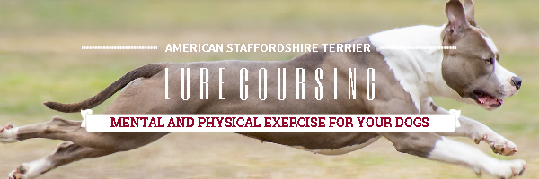
Lurecoursing.png
https://qldamstaffclub.com/images/FrontpageSlideshow/Lurecoursing.png
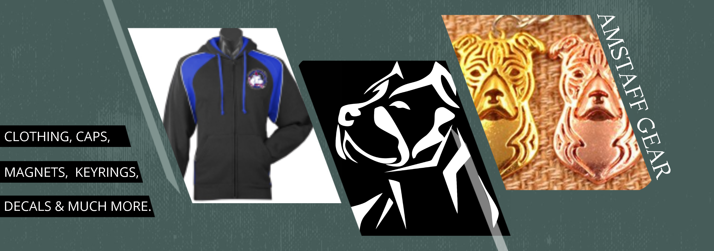
Promotion.png
https://qldamstaffclub.com/images/FrontpageSlideshow/Promotion.png
Merchandise.png
https://qldamstaffclub.com/images/FrontpageSlideshow/Merchandise.png
SHOWING.png
https://qldamstaffclub.com/images/FrontpageSlideshow/SHOWING.png
Shows.png
https://qldamstaffclub.com/images/FrontpageSlideshow/Shows.png
Versatile.png
https://qldamstaffclub.com/images/FrontpageSlideshow/Versatile.png
Welcome!!
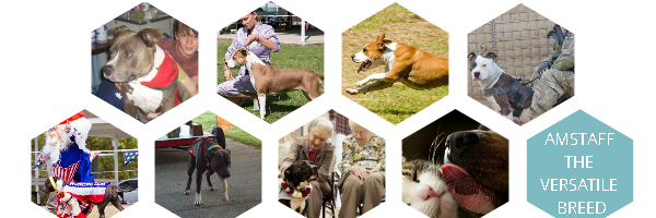
The American Staffordshire Terrier Club of QLD is dedicated to the preservation and promotion of the American Staffordshire Terrier.

2023 Club Sponsors!!


Sponsors
The K9 Professionals is one of Australia's largest dog training and behaviour consultancies, and also run Australia's largest online dog store.
Orivet Genetic Pet Care is a leading genetic testing organisation offering an extensive range of personalised genetic services for companion animals.
ShowManager - Online Entry and Dog Show management system
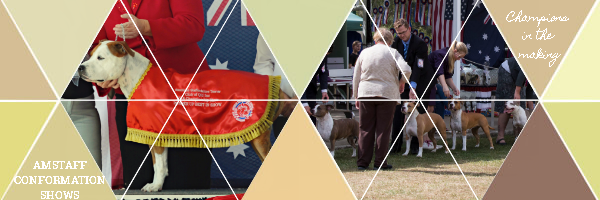
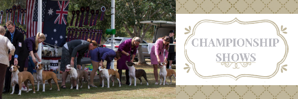
The ASTCQ Inc hold a range of events for all enthuasists. Conformation Shows, Lure Coursing, Weight Pull to name a few.

The ASTCQ have a Orivet Authorised DNA Collector available at all events, if you need DNA collected, please contact Karen via text on 0422 425 413.
Health
Cerebellar Ataxia (NCL-A) in Amstaffs
The American Staffordshire Terrier is a relatively healthy breed, though it is affected by some unpleasant disorders. One of the most serious disorders from which this breed suffers is called Hereditary Ataxia or Cerebellar Ataxia. This is a neurological disorder of a serious nature and as of yet there is no cure; it seems as if the gene governing the disorder is quite widespread in the breed and often avoiding the breeding of affected dogs is difficult due to the late onset of symptoms. Research to find out the exact mode of inheritance and to find some kind of treatment is ongoing and owners of dogs with the disease are encouraged to allow their dogs to participate in trials and studies.
This condition involves the cerebellum, a very important part of the brain; it’s located at the base of the brain, just above the brain stem and spinal cord. The most important function of the cerebellum is the coordination of movement. It takes information regarding the position of the body and its muscles and integrates it with pathways coming from higher structures of the brain that involve movement commands. Based on the body’s position, the cerebellum is able to control movements with a high level of precision; the cerebellum is constantly working to maintain posture and muscle tone. If this area is damaged, the individual will have great difficulty moving and often swaggers and sways; there may even be cognitive damage as well.
Hereditary ataxia is a condition in which there is widespread degeneration of the cerebellum. While the signs resulting from hereditary ataxia could be indicative of a wide variety of diseases or problems that affect the cerebellum, this particular disease is characterized by a certain order and rate of appearance of signs. First, you’ll see your dog behave somewhat clumsily and he could begin to sway occasionally. Clumsiness worsens over time with the progression of the disease and soon the dog will constantly fall over, losing his balance. You may also notice rapid eye and head movements and walking will become much more difficult; weight loss is often seen in dogs suffering from hereditary ataxia. These symptoms usually do not occur in dogs younger than two years of age.
Exactly how the disease develops is still poorly understood and it actually seems to be different in different dogs. Signs are slow to develop in some dogs, while they develop rapidly in other dogs. As of now, this is a fatal disease as there is no cure and a normal life is impossible; Amstaffs with hereditary ataxia are more often than not humanely euthanized by the time they are 7 or 8 years old. There are no diagnostic tests at the moment to specifically detect hereditary ataxia; confirmation of the presence of the disease comes only through an autopsy, after death. If you see your Amstaff exhibiting strange signs, you must immediately take the dog to a veterinary neurologist who will perform a wide variety of tests to rule out any other possible condition that could lead to the same signs.
Cerebellar Ataxia is inherited as a autosomal recessive trait, which means a dog must have two defective copies (one from each parent) to be affected. A dog once tested will be classified as one of the following three classifications:
Clear - 2 normal copies of the implicated gene - the dog does not have ataxia, is not a carrier for ataxia and cannot produce carrier or affected offspring.
Carrier - 1 normal copy and one defective copy of the implicated gene - the dog is not affected by ataxia but is a carrier of the gene. He can pass the gene onto his offspring.
Affected - 2 defective copies of the implicated gene - the dog has ataxia, and will pass the gene onto offspring.
The approximate breakdown rate of breeding a clear, carrier and affected dog is as follows:

It should be noted if 2 Clear dogs are bred together, the resulting pups are clear by parentage and do not require testing. If you are purchasing a Amstaff pup, please ask the status of the parents Ataxia test results. Clear by parentage can only be used when the pups are DNA parentaged to the pups, without the DNA parentage certificate, clear by parentage should not be used.
A test for Cerebellar Ataxia was found in 2008, there is no reason for a breeder not to have their dogs tested.
To have your dog tested, please visit Antagene's website. You will need to request a DNA Swab kit from Antagene, once it arrives, take your dog and the test kit to your vet. Your dog will need to be microchipped. The vet will do a non invasive cheek swab and sign all the paperwork for Antagene, send the form and swab back to them along with payment and a few weeks later you will receive a certificate with your dogs status!
Hip dysplasia. These two words terrify dog breeders and owners, but the truth is hip dysplasia can happen to any size dog. This condition can drastically reduce a dog’s quality of life and is painful for owners to watch. The good news is that embracing the responsibilities of owning a breed at risk, and educating yourself about potential health conditions, such as hip dysplasia, can go a long way toward keeping your dog comfortable.
Here is what all dog owners should know about hip dysplasia, including the symptoms, treatment options, and preventative measures you can take to keep your dog healthy, happy, and active.
What Is Canine Hip Dysplasia?
Canine hip dysplasia is a common skeletal condition, more common in large or giant breed dogs, although it can occur in smaller breeds, as well. To understand how the disease works, owners first need to understand the basic anatomy of the hip joint.
The hip joint functions as a ball and socket. In dogs with hip dysplasia, the ball and socket do not fit or develop properly, rubbing and grinding instead of sliding smoothly. This results in deterioration over time and an eventual loss of function of the joint itself.
What Causes Hip Dysplasia in Dogs?
Several factors lead to the development of hip dysplasia in dogs, beginning with genetics. Factors such as excessive growth rate, types of exercise, and improper weight and nutrition can magnify this genetic predisposition.
Correct nutrition as youngsters is very important to all puppies. These foods help prevent excessive growth, which can lead to skeletal disorders such as hip dysplasia, along with elbow dysplasia and other joint conditions. Slowing down these breeds’ growth allows their joints to develop without putting too much strain on them, helping to prevent problems down the line.
Improper nutrition can also influence a dog’s likelihood of developing hip dysplasia, as can too much exercise – or too little. Obesity puts a lot of stress on your dog’s joints, which can exacerbate a pre-existing condition such as hip dysplasia or even cause hip dysplasia.
Symptoms of Hip Dysplasia in Dogs
Some dogs begin to show signs of hip dysplasia when they are as young as four months of age, while other dogs develop it in conjunction with osteoarthritis as they age. In both cases, there are quite a few symptoms associated with hip dysplasia that larger breed dog owners should be familiar with. These symptoms may vary depending on the severity of the disease, the level of inflammation, the degree of looseness in the joint, and how long the dog has suffered from hip dysplasia.
- Decreased activity
- Decreased range of motion
- Difficulty or reluctance rising, jumping, running, or climbing stairs
- Lameness in the hind end
- Looseness in the joint
- Narrow stance
- Swaying, “bunny hopping” gait
- Grating in the joint during movement
- Loss of thigh muscle mass
- Noticeable enlargement of the shoulder muscles as they compensate for the hind end
- Pain
- Stiffness
- Diagnosing Hip Dysplasia in Dogs
At your dog’s regular checkup, your veterinarian will perform a physical exam. Sometimes this exam is enough for your veterinarian to suspect hip dysplasia. In other cases, it is up to you, as the owner, to let your veterinarian know that your dog is experiencing discomfort.
If you or your veterinarian suspect hip dysplasia, one of the first things that your veterinarian will do is manipulate your dog’s hind legs to test the looseness of the joint and to check for any grinding, pain, or reduced range of motion. Your dog’s physical exam may include blood work because inflammation due to joint disease can be indicated in the complete blood count. Your veterinarian will also need a history of your dog’s health and symptoms, any possible incidents or injuries that may have contributed to these symptoms, and any information you have about your dog’s parentage.
The definitive diagnosis usually comes with a radiograph (x-ray). Your veterinarian will take radiographs of your dog’s hips to determine the degree and severity of the hip dysplasia, which will help determine the best course of treatment for your dog.
Treating Hip Dysplasia in Dogs
There are quite a few treatment options for hip dysplasia in dogs, ranging from lifestyle modifications to surgery.
If your dog’s hip dysplasia is not severe, or if your dog is not a candidate for surgery for medical or financial reasons, your veterinarian may recommend a nonsurgical approach. Depending on your dog’s case, the vet may suggest the following:
- Weight reduction to take stress off the hips
- Exercise restriction, especially on hard surfaces
- Physical therapy
- Anti-inflammatory medications (nonsteroidal anti-inflammatory drugs (NSAIDS), aspirin, corticosteroids)
- Joint fluid modifiers
If your dog is a good candidate for surgery, there are more options. While there are quite a few different surgical strategies, the most common surgeries veterinarians use to treat hip dysplasia in dogs are:
- Double or triple pelvic osteotomy (DPO/TPO)
- Femoral head ostectomy (FHO)
- Total hip replacement (THR)
DPO/TPO surgery is usually performed in young dogs less than 10 months old. In this surgery, the surgeon improves the function of the ball and socket joint by selectively cutting the pelvic bone and rotating the segments.
FHO surgery can be performed on young and mature dogs. The surgery involves cutting off the femoral head, or “ball,” of the hip joint, which results in the body creating a “false” joint that reduces the discomfort associated with hip dysplasia. FHO does not recreate normal hip function, but it can be a successful pain management strategy.
THR - The most effective surgical treatment for hip dysplasia in dogs is a total hip replacement. The surgeon replaces the entire joint with metal and plastic implants, returning hip function to a more normal range and eliminating most of the discomfort associated with hip dysplasia.
Preventing Hip Dysplasia in Dogs
Not all cases of hip dysplasia can be prevented, but there are some steps you can take to reduce your dog’s risk of developing this disease.
Keeping your dog’s skeletal system healthy should start when your dog is young. Feeding your puppy an appropriate diet, especially if you have a large breed puppy, will give her a head start on healthy bone and joint development and help prevent the excessive growth that leads to the disease.
As your dog grows, providing her with appropriate levels of exercise and a healthy diet will prevent obesity, which is a major contributing factor to hip dysplasia. Obesity also causes many other health problems in dogs, from diabetes to elbow dysplasia, so hold off on the table scraps and other fatty foods.
As a prospective purchaser of a new dog, do your research on the breed of your choice and find a responsible breeder that does the appropriate health screenings for that breed, such as radiographs for hip dysplasia, elbow dysplasia, etc.
Prognosis for Dogs With Hip Dysplasia
Dogs with hip dysplasia often lead long, full lives, especially with treatment. If you think that your dog has hip dysplasia, or if your dog has recently been diagnosed with hip dysplasia, talk to your veterinarian about the treatment options and lifestyle changes you can make to keep your dog comfortable well into old age.

What does the grading all mean and the equilivant in different countries?
| OFA (USA) |
FCI (European) | BVA (UK/ Australia) |
SV (Germany) |
| E (Excellent) | A-1 | 0-4 (no > 3/hip) | Normal |
| G (Good) | A-2 | 5-10 (no > 6/hip) | Normal |
| F (Fair) | B-1 | 11-18 | Normal |
| B (Borderline) | B-2 | 19-25 | Fast Normal |
| M (Mild) | C | 26-35 | Noch Zugelassen |
| Mod (Moderate) | D | 36-50 | Mittlere |
| S (Severe) | E | 51-106 | Schwere |
The common heart diseases in Amstaffs are as follows:

The Normal Heart
Patent ductus arteriosus (PDA):
The problem is the failure of the ductus arteriosis to close shortly after birth and thereby allows continued flow of blood between the aorta and pulmonary artery. This was originally though to be the most frequently encountered congenital cardiac defect in all dogs. It is now believed by cardiologist that aortic stenosis is the most common.
Usually this condition presents itself at 6 to 12 weeks of age and is more common in females. Signs of heart failure are usually absent. Often a prominent pulse may be noted in many unusual areas on the body (ie.gums). A continuous murmur is present.
Prognosis: Treatment for PDA is via surgical correction or coil occlusion. In the hands of a skilled surgeon the survival rate is 90%. Without surgery, it is estimated, though not substantiated, that 60% will die by 1 year.

Aortic Stenosis (AS):
AS is the most common congenital cardiac defect and most difficult to diagnose in the dog. The stenotic lesion may occur in the subvalvular position, valvular position or supravalvular position. The subvalvular is the most common in dogs.
This has been documented in many breeds, including Bull Terriers. It should be noted this is the most common condition that causes the exploding hearts in Amstaffs. The genetic factor(s) of AS are not known as of yet. It is believed to be polygenetic, and therefore very difficult to eliminate from the gene pool. Only through the testing of all breeding stock and strict culling of positive animals and producers of positive animals are there hopes to eventually eliminate this condition.
AS can be present at birth or develop up to one year of age. It has not been documented to develop in dogs over 1 year. It can be present as adults with an incidental murmur. Signs of heart failure are rare. Dysrhythmias with pulse deficits may be noted. Systolic heart murmur is located over left heart base and radiates of the neck and to the right hemothorax.
Definitive diagnosis can be done via Doppler echocardiography or Cardiac catheterization. EKG will show normal and diagnosis for AS is inconclusive. A presumptive diagnosis can be done via ausculation of a left systolic heart murmur with a weak femoral arterial pulse.
Treatment includes beta blockers (though they are controversial) or very limited surgical options. Though heart failure is rare, it is more likely to occur in cases of moderate and severe AS if concurrent mitral valve insufficiency is present and severe.
Prognosis:
· Most cases of AS of marked severity result in sudden death.
· Mild cases of AS live a full life (both normal quality and duration)
· Even cases of moderate stenosis can anticipate a life of normal length and quality.
· Work with Doppler echocardiography has revealed that if the velocity of flow across the left ventricular outflow is less than 4 m/s by the time the dog is mature, it appears that the dogs can anticipate a life of normal length and quality. If the velocity is greater than 5 m/s it will likely succumb to this disorder.
· Progression of AS is very slow in mature dogs.
· It has been reported that if dogs survive with AS beyond 3 years, they usually do not have AS severe enough to produce a marked effect on left valve performance.

Pulmonary Stenosis (PS):
PS is the third most prevalent cardiac disorder in the dog. A stenotic lesion may occur in subvalvular, valvular or supravalvular position. The valvular disorder is the most common in dogs. Symptoms include fatigue and exercise intolerance. A definitive diagnosis for PS is via the Doppler echocardiography or Cardiac catherization test. EKG provides insight into the disorder. A presumptive diagnosis can be made via ausculation of a left basilar systolic heart murmur with a normal femoral arterial pulse. Dilation of the main pulmonary artery on thoracic radiographs may be seen.
The treatment of choice for severe PS is surgical correction. However the efficacy of surgical intervention remains to be substantiated. Medication to control dysrhythmias and fluid around the heart may also be an option.
Prognosis: Mild cases can be expected to live a full life with this abnormality. Severe cases of PS may die suddenly or develop signs of right heart failure and die in 6-12 months.

Ventricular Septal Defect (VSD):
This abnormality is a defect allowing blood flow between the left and right ventricle. This defect usually presents itself in puppies. A large defect will stunt growth and the pup will fail to thrive.
A definitive diagnostic test for VSD is via Doppler or Cardiac catheterization. A presumptive diagnosis can be made by ausculation of a right sternal border systolic heart murmur or radiographic evidence of pulmonary overcirculation.
Symptoms include reduced exercise tolerance, fatigue, possible heart murmur and jugular distention.
Treatment for mild VSD requires no therapy, whereas severe VSD requires surgical repair. Prognosis for severe VSD if pulmonary hypertension develops is very poor.

Mitral Valve Dysplasia (MVD):
The most common disorder of the mitral valve is the partial backflow (regurgitation) of blood through the valve. Regurgitation is usually the result of valvular degeneration, which occurs most often in older members of the smaller dog breeds. Severe mitral regurgitation, not only produces significant increases in the left side of the heart, it is frequently accompanied by varying degrees of congestive heart failure.

Tricuspid Valve Dysplasia (TVD):
This valve is located between the right atrium and the right ventricle of the heart. The purpose of this valve is to control the backflow of blood during contractions of the heart.
During normal fetal development, the tricuspid valve flaps are adhered to the wall separating atrium from ventricle. As the fetal development progresses under normal circumstances the adhesive bonds holding the valve open will degenerate, allowing the valve flaps to move into their proper position.
One of the primary causes of TVD is the failure of the bond to degenerate. Depending on the severity of the malformation, the work on the right side of the heart increases.
Symptoms of TVD are dependent upon the extent of the malformation, but some of the most common symptoms re: fluid retention, cool extremities and exercise intolerance (possibly followed by collapse).
TVD in dogs is usually congenital (present at birth). Due to the fact that this condition (when it occurs) appears in several littermates, and tends to be more prevalent in some family bloodlines than others, it is suspected that the tendency to have this birth defect is hereditary. It is hoped that through screening of breeding stock and their lineage (parents, grandparents, littermates, aunts, uncles, etc) efforts can be made to eliminate susceptible bloodlines from breeding programs.

Elbow dysplasia is a general term used to identify an inherited polygenic disease in the elbow of dogs. Three specific etiologies make up this disease and they can occur independently or in conjunction with one another. These etiologies include:
- Pathology involving the medial coronoid of the ulna (FCP)
- Osteochondritis of the medial humeral condyle in the elbow joint (OCD)
- Ununited anconeal process (UAP)
Studies have shown the inherited polygenic traits causing these etiologies are independent of one another. Clinical signs involve lameness which may remain subtle for long periods of time. No one can predict at what age lameness will occur in a dog due to a large number of genetic and environmental factors such as degree of severity of changes, rate of weight gain, amount of exercise, etc. Subtle changes in gait may be characterized by excessive inward deviation of the paw which raises the outside of the paw so that it receives less weight and distributes more mechanical weight on the outside (lateral) aspect of the elbow joint away from the lesions located on the inside of the joint. Range of motion in the elbow is also decreased.
Fractured Coronoid Process (FCP)
Who is usually affected?
-Young dogs of large to giant breeds
-Most frequently affected breeds include Labrador retrievers, Golden retrievers, Rottweilers, and Bernese mountain dogs
What is happening?
-The 3 bones of the elbow joint fit poorly, causing abnormal pressure on the ulna
-A small piece of bone associated with the ulna in the front of the joint (coronoid process) breaks off
-Swelling and pain result from the altered joint mechanics and cartilage destruction
-Arthritis develops
Clinical signs you might notice in your pet
-Limping on a front leg after rest or exercise
-Tiring easily with play
-Resting more than other dogs of similar age and breed, "mellow" puppy
-Head bobbing during walking or running
-Sitting or standing crookedly with a front leg turned outward
Diagnosis
-Careful orthopedic examination to determine which joint(s) are affected
-X-rays are used to evaluate the condition of joint surfaces
-CT scanning may be useful in select cases to evaluate joint surfaces
Surgical treatment
-Removal of the bone fragment and any damaged cartilage (curettage)
-Surgery through small portals (arthroscopy) or a larger incision (arthrotomy) may be indicated
-Surgery on both elbows may be recommended, as the disease is frequently present on both sides
Special postoperative care
-Patient activity is generally limited for 4-6 weeks following surgery, allowing time for joint swelling to subside
Expected results after surgery
-Much initial improvement in the degree of pain and limping
-Ultimate outcome depends on the amount of joint damage present prior to surgery and the degree of elbow misfit ( incongruity )
-Moderate exercise, weight control and medication may be recommended for the long term management of optimal joint health.
Osteochondritis Dissecans (OCD)
Who is usually affected?
-Young dogs of large to giant breeds
What is happening?
-Abnormal maturation of the bone that supports cartilage within joints leads to cartilage thickening, cracking, and exposure of the underlying bone
-Swelling and pain result from the altered joint mechanics and cartilage destruction
-Arthritis develops
-Most commonly affected joints are the shoulder, elbow, knee (stifle), and ankle (hock)
Clinical signs you might notice in your pet
-Limping after rest or exercise
-Tiring easily with play
-Resting more than other dogs of similar age and breed, "mellow" puppy
-Head bobbing during walking or running
-Sitting crookedly with one leg held outward (though this can be normal for puppies)
Diagnosis
-Careful orthopedic examination to determine which joint(s) are affected
-X-rays are used to evaluate the condition of joint surfaces
-X-ray with a contrast liquid (arthrogram) may be needed to fully evaluate the shoulder joint
Surgical treatment
-Removal of the damaged cartilage (curettage)
-Surgery through small portals (arthroscopy) or a larger incision (arthrotomy) may be indicated
Special postoperative care
-Patient activity is generally limited for 4-6 weeks following surgery, allowing time for joint swelling to subside
Expected results after surgery
-Shoulder: Excellent results (pain relief and gradual return to a normal gait) are expected due to the relatively small area of cartilage usually involved and the loose fitting mechanical nature of the joint
-Elbow: Good results are expected unless the area of cartilage involved is very large
-Stifle: Good results are expected if the area of cartilage involved is in a non-weight bearing portion of the stifle; variable results are expected if the area of cartilage involved is in a weight bearing portion of the stifle (additional surgery may be indicated to decrease weight bearing on this site)
-Hock: Fair to poor results (incomplete pain relief and retention of some abnormal gait) are expected due to the relatively large area of cartilage usually involved and the tight fitting mechanical nature of the joint
-Moderate exercise, weight control and medication may be recommended for the long term management of optimal joint health.
Ununited Anconeal Process (UAP)
What is happening?
-The 3 bones of the elbow joint fit poorly, causing abnormal pressure on the ulna
-A small piece of bone associated with the ulna in the back of the joint (anconeal process) fails to attach to or breaks off of the ulna
-Swelling and pain result from the altered joint mechanics and cartilage destruction
-Arthritis develops
Clinical signs you might notice in your pet
-Limping on a front leg after rest or exercise
-Tiring easily with play
-Resting more than other dogs of similar age and breed, "mellow" puppy
-Head bobbing during walking or running
-Sitting or standing crookedly with a front leg turned outward
Diagnosis
-Careful orthopedic examination to determine which
joint(s) are affected
-X-rays are used to evaluate the condition of joint surfaces
Surgical treatment
-Depending on the specifics of each case, recommended surgical options may include:
-Removing the loose piece of bone
-Relieving the pressure on the ulna by cutting the bone (osteotomy) below the elbow joint, encouraging the previously loose piece of bone (anconeal process) to attach to the ulna
Special postoperative care
-Patient activity is generally limited for 4-6 weeks following surgery, allowing time for joint swelling to subside
-Patient activity may be limited for a longer period of time (6-12 weeks) with ulnar osteotomy, allowing time for the anconeal process to fuse with the ulna
Expected results after surgery
-Much initial improvement in the degree of pain and limping
-Ultimate outcome depends on the amount of joint damage present, prior to surgery and the degree of elbow misfit (incongruity)
-Moderate exercise, weight control and medication may be recommended for the long term management of optimal joint health.

Signs and Symptoms
Dog hyperthyroidism is an endocrine disease that causes overproduction of the hormone thyroxin. The presence of excessive amounts of this hormone then affects the animal's entire system, causing many problems. While this condition is quite rare in dogs and is far more prevalent in cats, it does occasionally affect canines. If you're concerned that your canine companion may be suffering from hyperthyroidism, there are a number of characteristic warning signs that you can look out for.
One of the most common and most noticeable symptoms of canine hyperthyroidism is weight loss despite an unusually ravenous appetite. If your pet is eating more than normal and yet seems to be getting thinner and thinner, you should have him or her checked out for this endocrine condition. Sometimes dogs with this disease will eat so quickly that they end up vomiting. Some pets may also experience diarrhea. Also, excessive thirst may accompany the characteristic voracious appetite.
Other very common warning signs of this disease include hyperactivity and nervousness. Your pet may become more active and restless than normal and may also show signs of irritability. These symptoms are often accompanied by an elevated heart rate. In addition to beating faster, an afflicted dog's heart may also exhibit stronger beats, which you may be able to feel by placing your hand against your dog's chest. Some dogs that are suffering from hyperthyroidism also tend to breathe more rapidly than usual.
At times, a canine with this endocrine disease will also experience changes in its coat. For example, a dog's fur may become dull and appear unusually unkempt. Thus, if you notice that your pet is displaying any of the above symptoms, he or she may be suffering from the production of too much thyroxin that is caused by hyperthyroidism. If you suspect that your dog may indeed be suffering from this condition, it's important to contact your veterinarian as soon as possible.
Diagnosis
When a case of canine hyperthyroidism is suspected, a veterinarian will make a diagnosis based upon clinical signs and blood tests. First of all, the doctor will speak to you as the pet owner about your dog's behavior at home and the warning signs that you have observed. Then the practitioner will conduct a physical exam, checking for symptoms such as an enlarged thyroid and an elevated heart rate. These steps will then be followed by blood tests.
General blood tests will show whether or not there is a stress response in your pet. In such cases, the blood sample may show elevated levels of red blood cells and some white blood cells. At the same time, certain types of white blood cells -- lymphocytes and esinophils -- will appear in lower amounts than normal. Furthermore, certain other substances such as the liver enzyme ALT, glucose, phosphorus, and creatine may also appear in high levels due to complications in other parts of the body that are caused by the hyperthyroidism.
More specifically, a blood test will be conducted to check for levels of the thyroid hormone known as T4. This is a simple technique that is relatively inexpensive; however, it is not always reliable as the results can appear falsely low due to the effects of medication or other illness. As a result, another technique known as Free T4 or FT4 testing may be required. This test is much more reliable and is far more reflective of the thyroid's true state and function. This method of testing is more expensive than basic T4 testing, but it may be required in order to make a confident diagnosis of hyperthyroidism in your pet.
On some occasions, imaging techniques such as ultrasound may be employed by a veterinarian. Ultrasound allows a doctor to examine the physical characteristics of the canine patient's thyroid, revealing masses or other abnormalities. However, blood tests and clinical signs are the most common avenues used to arrive at a diagnosis of canine hyperthyroidism.
Pathophysiology
The thyroid is one of the major endocrine glands in the canine body. It is a small, double-lobed structure located in the neck that is responsible for producing the hormones thyroxine (T4) and triiodothyronine (T3). These hormones play a role in several physiological systems and functions, including the regulation of metabolism. When hyperthyroidism occurs, this gland becomes overactive and produces higher than normal amounts of thyroid hormones. Most often this overproduction affects the hormone T4, but sometimes it causes elevated levels of T3 or both hormones together.
Since thyroid hormones are involved in numerous physiological functions and pathways, many parts of the body are affected when normal levels of these hormones are not maintained. In fact, since thyroid hormones play an important role in metabolism, hyperthyroidism has an effect on every single cell in the body. This is because every cell requires and burns fuel. However, certain systems and functions are affected more noticeably than others, giving rise to the characteristic symptoms displayed by animals suffering from this endocrine disorder. For example, the increased levels of thyroxine intensify a dog's metabolism, which leads to the weight loss and ravenous appetite that an afflicted animal experiences.
In fact, this condition causes the intensification of many functions in the canine body. In addition to increased metabolism, the high levels of thyroxine will also cause an increased heart rate and higher blood pressure than normal. If this disease goes untreated, these changes can eventually cause the animal's heart to grow in size which leads to a condition known as myocardial hypertrophy. This occurs because the dog's heart is attempting to respond to the increased stress and pressure being exerted upon this organ. Understandably, this secondary condition can be quite problematic, eventually causing cardiac dysfunction.
As a result, it's clear that hyperthyroidism can have serious effects upon many parts of a dog's system. No cell is left untouched by this disease that is characterized by over activity on the part of a canine's thyroid.
Causes
While hyperthyroidism may be caused by a variety of factors in humans, cats, and other animals, in dogs it is almost always caused by cancer of the thyroid, or thyroid carcinoma. This disease involves the growth of a cancerous nodule or other mass on the thyroid gland. This type of malignant mass then interferes with the proper function of the thyroid, causing hyperthyroidism and the overproduction of hormones. Nearly all cases of hyperthyroidism in canines are caused by this type of cancer. Thus, if your dog is suffering from overproduction of thyroid hormones due to this endocrine disorder, it's very likely that thyroid carcinoma is at the root of the problem.
Treatment When a dog is suffering from hyperthyroidism, there are three traditional methods of treatment. These include medication, surgery, and radioactive iodine treatment. In the case of medication, daily doses of specific pharmaceuticals can help to prevent the overproduction of thyroid hormones that this disease causes. In most cases, normal levels of hormone production can be reached in approximately one to three weeks. Depending upon your dog's preferences and needs, this type of medication can often be administered in different forms such as in a solid tablet, in liquid form, or as a gel that can be rubbed into the animal's ears.
In some situations, surgery may be required to treat this condition. This technique can involve the removal of either part of the thyroid or the entire gland. Radioiodine treatment is another possibility. This method uses radioactive iodine to kill the overactive cells of the thyroid. While this is considered one of the most effective treatments for hyperthyroidism, not all animals are good candidates for the procedure and a lengthy hospitalization is often required.
In addition to these traditional treatments, some homeopathic remedies can be helpful with regards to calming an overactive thyroid and reducing the symptoms of this disorder. Numerous natural substances have properties that are very beneficial in this respect. For example, bungleweed has properties which inhibit thyroid stimulating hormones, reducing the overactive production of thyroxine and triiodothyronine. This natural substance also relieves the irritability that can sometimes result from hyperthyroidism. It also helps to strengthen certain heart functions, as does hawthorn. Other useful homeopathic ingredients include lemon balm and valerian. These substances relax spasms, relieve tension, and reduce excitability. Homeopathic products geared towards treating canine hyperthyroidism may contain a combination of these ingredients, as well as other natural ingredients which have a beneficial effect upon an overactive thyroid.
If your dog is suffering from unusual symptoms, the most important thing that you can do is to make sure that he or she is examined by a veterinarian immediately. If a diagnosis of canine hyperthyroidism is made, the veterinary doctor can help you to select and implement the most appropriate treatment option for your individual pet.

Quick Links
Club History
The American Staffordshire Terrier Club of QLD was created as a Social Club in 2002. In 2006, the Club became Affiliated with DogsQLD, allowing the Club to hold Shows and Sporting Events.
Buying an Amstaff?

Breed Specific Legislation


BSL
hqdefault.jpg
8506419.jpg
BSL.jpg
2013-06-23_21.48.26.jpg
https://qldamstaffclub.com/images/BSL/2013-06-23_21.48.26.jpg
BSL1.jpg
image_thumb_6.png
stop_bsl_poster.jpg
Amstaff Rescues in Australia


Rescues
QLD Staffy & Amstaff Rescue
We are all about improving the lives of Staffordshire Bull Terriers, American Staffordshire Terriers and their crosses in need of help.
Staffies United Rescue
Staffies United Rescue (SUR) is a not for profit dog rescue. We are entirely managed and run by volunteers. We are dedicated to helping Staffies in need, improving the breed's reputation and we are highly committed to educating the community about responsible pet ownership.
Staffy Rescue
Staffy Rescue is a registered animal welfare charity committed to trying to save and rehome as many Staffordshire bull terriers and larger breed dogs as possible. Staffy Rescue rescues dogs from death row at pounds across NSW and kennels them at our shelter in north western Sydney.